Cover
Table of Contents
List of Illustrations
Maja Bondestam
Maria Kavvadia
Figure 1.1. Pirro Ligorio, Pyrrhichia saltatio [The Pyrrhic dance], 1573. Engraving from Girolamo Mercuriale, De arte gymnastica Libri sex, in quibus exercitationum omnium vetustarum genera, loca, modi, facultates & quidquid deniq. Ad corporis humani exer
Rosemary Moore
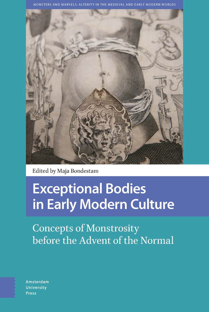
Figure 2.1. Detail of the monstrous creature in Johann Remmelin, ‘First Vision’, 1619. Etching and engraving from Catoptrum microcosmicum. Wellcome Library, London. Photograph: Rosemary Moore.
Figure 2.2. Johann Remmelin, ‘First Vision’, 1619. Etching and engraving from Catoptrum microcosmicum. Wellcome Library, London. Photograph: Rosemary Moore.
Figure 2.3. Johann Remmelin, ‘Second Vision’, 1619. Etching and engraving from Catoptrum microcosmicum. Wellcome Library, London. Photograph: Wellcome Library.
Figure 2.4. Johann Remmelin, ‘Third Vision’, 1619. Etching and engraving from Catoptrum microcosmicum. Wellcome Library, London. Photograph: Rosemary Moore.
Figure 2.5. Jacob Frölich, after Heinrich Vogtherr, Anatomy, or, a Faithful Reproduction of the Body of a Female, 1544. Woodcut. 55 x 25 cm. Wellcome Library, London. Photograph: Wellcome Library.
Figure 2.6. Detail of the Tetragrammaton in Johann Remmelin, ‘First Vision’, 1619. Etching and engraving from Catoptrum microcosmicum. Wellcome Library, London. Photograph: Rosemary Moore.
Figure 2.7. Detail of the devil in Johann Remmelin, ‘First Vision’, 1619. Etching and engraving from Catoptrum microcosmicum. Wellcome Library, London. Photograph: Rosemary Moore.
Figure 2.8. Detail of the fetus in Johann Remmelin, ‘First Vision’, 1619. Etching and engraving from Catoptrum microcosmicum. Wellcome Library, London. Photograph: Rosemary Moore.
Figure 2.9. Charles Estienne, Female anatomical model showing the location of the caesarean cut, 1545. Woodcut from p. 260 of Charles Estienne, De dissectione partium corporis humani [On the dissection of the parts of the human body] (Parisiis: Apud Simon
Figure 2.10. Detail of the main anatomical figures with the flaps raised. From Johann Remmelin, ‘First Vision’, 1619. Etching and engraving from Catoptrum microcosmicum. Wellcome Library, London. Photograph: Rosemary Moore.
Pablo García Piñar
Cécile Tresfels
Parker Cotton
Maja Bondestam
Figure 6.1. Johannes Schefferus, A boy with a so-called prodigious appearance. Drawing from Chapter IV of Johannes Schefferus, ‘Variae historiae’, 1670-1679, Uppsala University Library, MS X 292. Photograph: Uppsala University Library.
Figure 6.2. Johannes Schefferus, The monster from Lillebered. Drawing from Chapter VIII of Johannes Schefferus, ‘Variae historiae’, 1670-1679, Uppsala University Library, MS X 292. Photograph: Uppsala University Library.
Figure 6.3. Johannes Schefferus, A large stone, found inside the bladder of a man. Drawing from Chapter III of Johannes Schefferus, ‘Variae historiae’, 1670-1679, Uppsala University Library, MS X 292. Photograph: Uppsala University Library.
Figure 6.4. Frontispiece, Julius Obsequens and Conrad Lycosthenes, Julii Obsequentis de prodigiis liber: cum annotationibus Joannis Schefferi […] accedunt Conr. Lycosthenis supplementum Obsequentis; item librorum à Scheffero editorum index (Stockholm, 167
Tove Paulsson Holmberg
Figure 7.1. Death and the infant: The child has a dull expression in its chubby face. Death squeezes it hard to her emaciated breast. She turns her back at us and increases her speed. Physician Lars Roberg, who inserted her portrait in Lijkrevningstavlor
Figure 7.2. Podalic version demands great mental and physical strength of the performer. He or she who wants to try it must “not stand sleeping, and have the vigor, and the heart, to carry it through”.27 Johan von Hoorn, Den Swenska Wäl-öfwade Jord-Gumman
Kathleen Long
Index